
Files in this item
Crystallization of Ranasmurfin, a blue coloured protein from Polypedates leucomystax
Item metadata
| dc.contributor.author | McMahon, Stephen | |
| dc.contributor.author | Walsh, MA | |
| dc.contributor.author | Ching, RTY | |
| dc.contributor.author | Carter, Lester | |
| dc.contributor.author | Dorward, M | |
| dc.contributor.author | Johnson, Kenneth Alan | |
| dc.contributor.author | Liu, Huanting | |
| dc.contributor.author | Oke, Muse | |
| dc.contributor.author | Block Jr, C | |
| dc.contributor.author | Kennedy, MW | |
| dc.contributor.author | Latiff, AA | |
| dc.contributor.author | Cooper, A | |
| dc.contributor.author | Taylor, Garry Lindsay | |
| dc.contributor.author | White, Malcolm Frederick | |
| dc.contributor.author | Naismith, James Henderson | |
| dc.date.accessioned | 2013-03-27T15:31:04Z | |
| dc.date.available | 2013-03-27T15:31:04Z | |
| dc.date.issued | 2006-11 | |
| dc.identifier | 367618 | |
| dc.identifier | 11e89212-83e4-4c0b-bd26-8a022a7ad069 | |
| dc.identifier | 000241681700017 | |
| dc.identifier | 33750576063 | |
| dc.identifier.citation | McMahon , S , Walsh , MA , Ching , RTY , Carter , L , Dorward , M , Johnson , K A , Liu , H , Oke , M , Block Jr , C , Kennedy , MW , Latiff , AA , Cooper , A , Taylor , G L , White , M F & Naismith , J H 2006 , ' Crystallization of Ranasmurfin, a blue coloured protein from Polypedates leucomystax ' , Acta Crystallographica. Section F, Structural biology and crystallization communications , vol. 62 , no. Pt 11 , pp. 1124-1126 . https://doi.org/10.1107/S1744309106040036 | en |
| dc.identifier.issn | 1744-3091 | |
| dc.identifier.other | ORCID: /0000-0003-1543-9342/work/47136132 | |
| dc.identifier.other | ORCID: /0000-0001-9486-566X/work/60428052 | |
| dc.identifier.uri | https://hdl.handle.net/10023/3437 | |
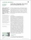
| dc.description.abstract | Ranasmurfin, a previously uncharacterized similar to 13 kDa blue protein found in the nests of the frog Polypedates leucomystax, has been purified and crystallized. The crystals are an intense blue colour and diffract to 1.51 angstrom with P2(1) symmetry and unit-cell parameters a = 40.9, b = 59.9, c = 45.0 angstrom, beta = 93.3 degrees. Self-rotation function analysis indicates the presence of a dimer in the asymmetric unit. Biochemical data suggest that the blue colour of the protein is related to dimer formation. Sequence data for the protein are incomplete, but thus far have identified no model for molecular replacement. A fluorescence scan shows a peak at 9.676 keV, indicating that the protein binds zinc and suggesting a route for structure solution. | |
| dc.format.extent | 3 | |
| dc.format.extent | 291534 | |
| dc.language.iso | eng | |
| dc.relation.ispartof | Acta Crystallographica. Section F, Structural biology and crystallization communications | en |
| dc.subject | QH426 Genetics | en |
| dc.subject.lcc | QH426 | en |
| dc.title | Crystallization of Ranasmurfin, a blue coloured protein from Polypedates leucomystax | en |
| dc.type | Journal article | en |
| dc.contributor.sponsor | BBSRC | en |
| dc.contributor.institution | University of St Andrews. School of Chemistry | en |
| dc.contributor.institution | University of St Andrews. Biomedical Sciences Research Complex | en |
| dc.contributor.institution | University of St Andrews. School of Biology | en |
| dc.contributor.institution | University of St Andrews. EaSTCHEM | en |
| dc.identifier.doi | 10.1107/S1744309106040036 | |
| dc.description.status | Peer reviewed | en |
| dc.identifier.url | http://www.scopus.com/inward/record.url?scp=33750576063&partnerID=8YFLogxK | en |
| dc.identifier.url | http://ukpmc.ac.uk/abstract/MED/17077494 | en |
| dc.identifier.grantnumber | BBS/B/14426 | en |
This item appears in the following Collection(s)
Items in the St Andrews Research Repository are protected by copyright, with all rights reserved, unless otherwise indicated.